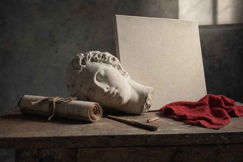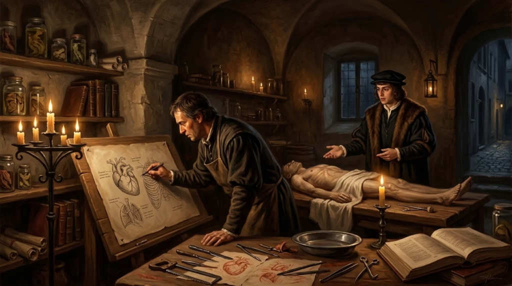
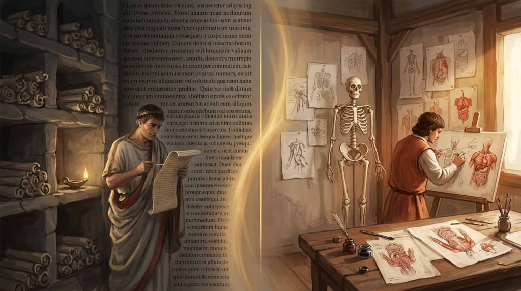
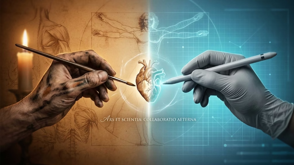
Picture a dimly lit room in early 16th-century Milan. Leonardo da Vinci bends over a dissection table, charcoal in hand, carefully rendering the intricate chambers of a human heart. Beside him stands Marcantonio della Torre, a young physician-anatomist, pointing out structures invisible to the untrained eye. Together, they’re creating something entirely new—illustrations that are simultaneously works of art and groundbreaking scientific discoveries.
This scene captures a remarkable moment in history when artists and scientists worked side by side, their disciplines not merely complementary but inseparable. The history of anatomical drawing isn’t just about medical progress or artistic achievement—it’s the story of a profound collaboration that revolutionized both fields and fundamentally changed how we understand the human body.
In this comprehensive guide, we’ll explore how this partnership emerged from the ashes of medieval restrictions, flourished during the Renaissance, and ultimately separated as medicine became more specialized. Drawing on museum collections, historical manuscripts, and scholarly research, we’ll discover what happened when science and art truly shared a studio.
Before the Shared Studio: Anatomy in the Ancient World
To understand why the Renaissance represented such a dramatic rebirth, we need to start where anatomical knowledge began—and where it was nearly lost.
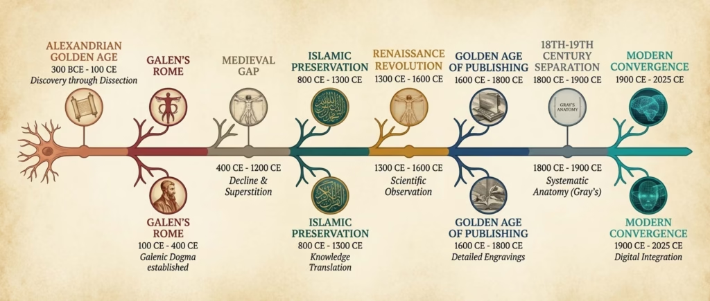
The Alexandrian Golden Age (300 BCE – 200 CE)
In the gleaming city of Alexandria, Egypt, something unprecedented was happening. For the first time in recorded history, physicians were systematically dissecting human bodies to understand how they worked.
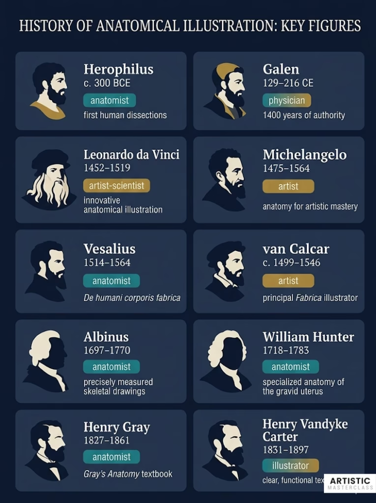
The city’s unique position made this possible. As a crossroads between Greek intellectual tradition and Egyptian mortuary practices, Alexandria developed a cultural openness to examining the dead. Under the patronage of the Ptolemaic dynasty, medical scholars like Herophilus and Erasistratus performed hundreds of dissections, making discoveries that wouldn’t be rediscovered for nearly two millennia.
Herophilus’s treatise On Anatomy synthesized findings from hundreds of human dissections. He distinguished between sensory and motor nerves, described the brain’s ventricles, and mapped the vascular system with remarkable accuracy. These thorough examinations were almost certainly accompanied by drawings, though frustratingly, none have survived. We know of their work only through later references and transcriptions.
When Rome conquered Alexandria, this golden age ended abruptly. Roman cultural attitudes viewed dissection as desecration, and the practice was forbidden throughout the empire.
Galen’s Legacy and Limitations
This is where Galen enters the story. Working in 2nd-century Rome as physician to gladiators and eventually to emperors, Galen faced an impossible situation. He believed passionately that direct observation was essential to understanding anatomy, but human dissection was strictly forbidden.
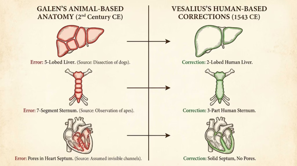
His solution was to dissect animals—particularly Barbary apes, which he considered “most like man”—and extrapolate to human anatomy. This approach led to genuine insights but also systematic errors that would mislead medicine for the next 1,400 years.
Galen described a liver with multiple lobes (true in dogs, not humans), claimed the human sternum had seven segments (true in apes), and incorrectly mapped blood flow through imagined pores in the heart’s septum. Yet his work was so comprehensive, so confidently presented, and so aligned with philosophical ideas about the body’s divine design that it became unquestionable dogma.
The Absence of Illustration
Here’s what’s crucial: this entire tradition remained almost entirely text-based. Ancient anatomical knowledge wasn’t illustrated because the visual culture simply didn’t exist to support it. Without the ability to reproduce images through printing, there was no practical way to standardize anatomical depictions across copies of manuscripts.
When Roman physicians like Celsus wrote about Alexandrian scholarship, they described what was seen—but couldn’t show it. This reliance on text alone meant that each generation interpreted descriptions differently, and critical visual details were lost in translation.
The Medieval Gap: When Text Replaced Observation
For roughly a thousand years, direct anatomical study nearly vanished from European medicine. Understanding this gap helps explain why the Renaissance breakthrough was so revolutionary.
European Restrictions and Symbolism
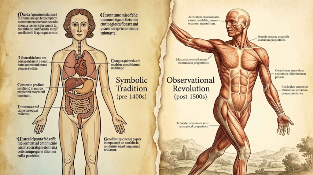
Medieval Christianity held the human body sacred. It was the vessel for the immortal soul, created in God’s image, destined for resurrection. Cutting it open was not just impractical—it was theologically problematic.
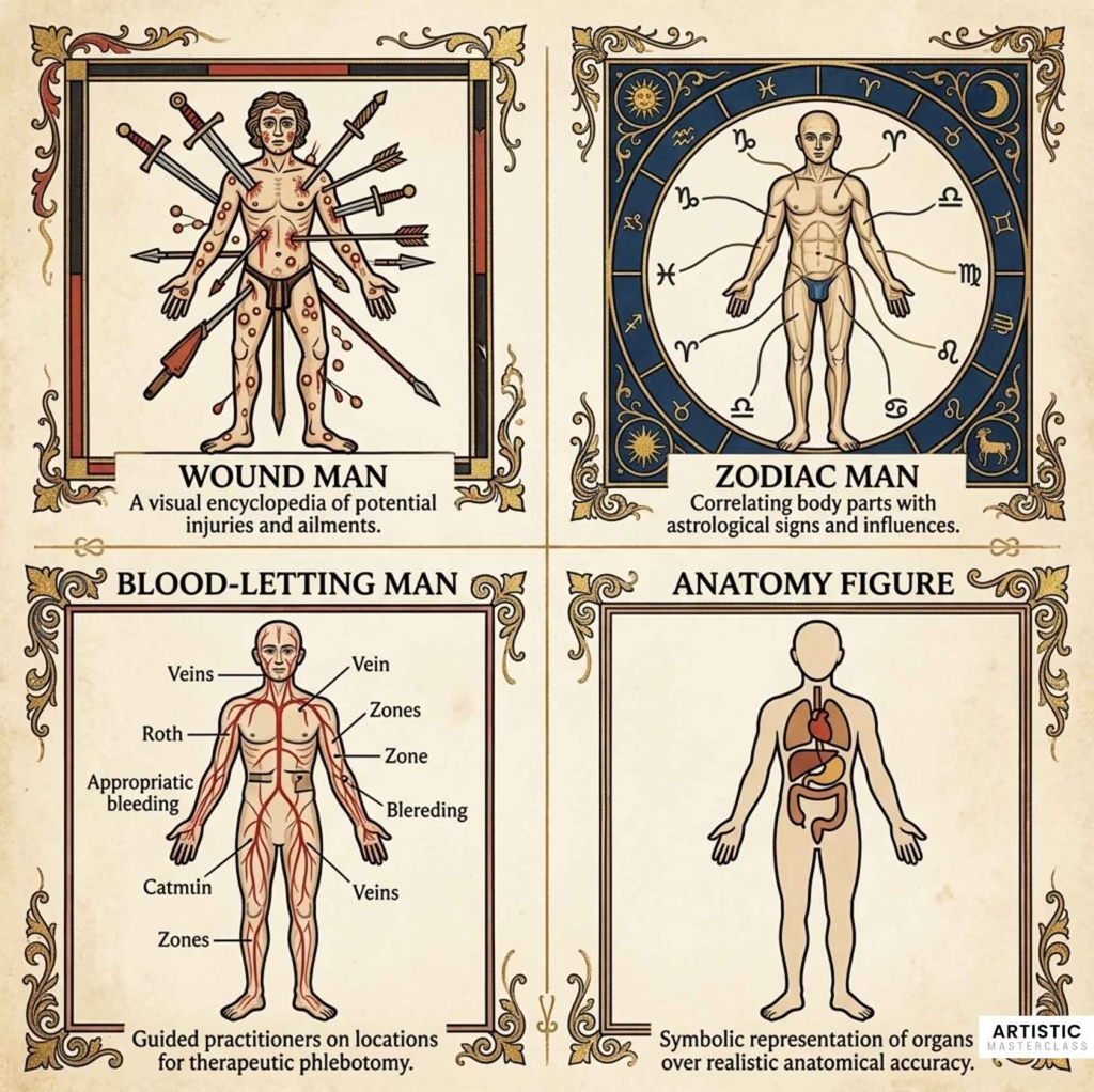
Medical knowledge came from reading ancient texts, particularly Galen, not from examining bodies. When medieval physicians needed visual references, they created symbolic figures that had little to do with actual anatomy.
The “Wound Man” showed a human figure pierced by various weapons and afflicted with diseases—a teaching tool about injuries, not anatomy. The “Zodiac Man” connected body parts to astrological signs for determining optimal treatment times. The “Blood-Letting Man” indicated where to open veins for therapeutic bleeding.
These images could be beautiful, even mesmerizing in their detail. But they represented medieval medicine’s focus on theory over observation, symbolism over structure.
Islamic Preservation and Innovation
While European anatomy stagnated, Islamic civilization was preserving and building upon classical knowledge. Scholars like Avicenna (Ibn Sina) systematically translated Greek and Roman medical texts into Arabic, adding their own observations and commentaries.
Mansur’s Anatomy, created in 14th-century Persia, contains some of the oldest surviving Islamic anatomical illustrations. The images show human figures with organs mapped in careful detail, demonstrating serious engagement with anatomical study despite Quranic restrictions on both human dissection and representational art.
Islamic physicians found creative solutions. Some performed limited dissections on war casualties. Others developed comparative anatomy through animal dissection. They preserved Galen’s errors along with his insights, but they also kept the flame of anatomical inquiry burning through Europe’s darkest centuries.
Late Medieval Awakening
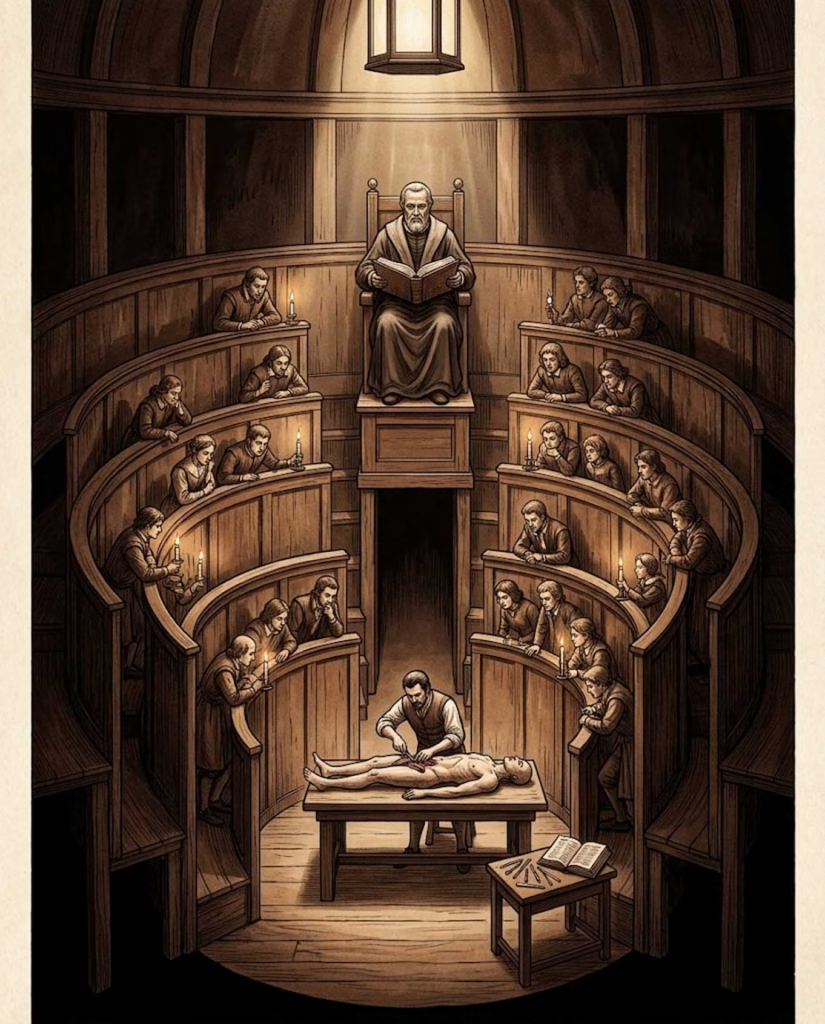
By the 13th century, European universities were beginning to reintroduce human dissection, though under strict limitations. At Bologna, Salerno, and later Padua, medical faculties received permission to dissect a small number of executed criminals each year.
These events were public spectacles. The anatomist, in full academic regalia, would sit in an elevated chair reading from Galen while a barber-surgeon performed the actual cutting. The goal wasn’t to discover new knowledge but to demonstrate the accuracy of ancient texts.
The first printed medical book with significant illustrations, Fasciculus Medicinae (1491), still showed this medieval character. Despite being printed during the Renaissance, its woodcuts depicted stylized figures that prioritized symbolism over observation.
But change was coming. The pieces were falling into place for a revolution.
The Renaissance Revolution: When Artists Became Anatomists
Multiple forces converged in 15th and 16th-century Italy to create an unprecedented collaboration between artists and scientists. Understanding why it happened there and then is crucial to understanding the transformation that followed.
The Perfect Storm: Why Renaissance Italy?
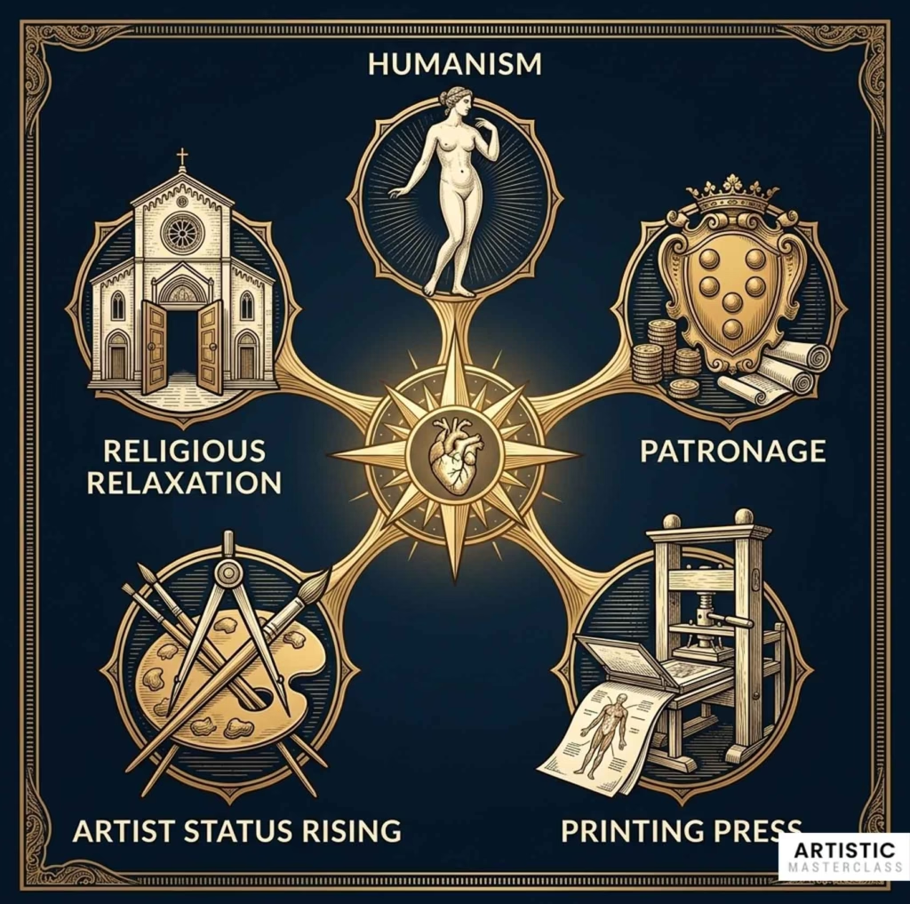
Humanism returned focus to human beings and the natural world. Classical texts that celebrated human form and rational inquiry were being rediscovered and newly translated. The human body wasn’t just a sacred vessel—it was worthy of study and celebration in its own right.
Wealth and patronage created an environment where both art and science could flourish. Families like the Medici in Florence didn’t just commission paintings and sculptures—they funded anatomical studies, collected scientific manuscripts, and brought together scholars and artists in their courts.
The printing press, perfected by Gutenberg around 1450, changed everything. For the first time, anatomical knowledge could be reproduced with identical illustrations across hundreds or thousands of copies. A drawing done well once could teach students across Europe.
Artist status was rising. No longer mere craftsmen, artists were becoming intellectuals. Leonardo da Vinci socialized with mathematicians and engineers. Michelangelo debated theology with cardinals. Artists were expected to understand geometry, perspective, optics—and anatomy.
Religious restrictions began to relax, carefully and selectively. The Church still controlled dissection, but university faculties could obtain permission for limited studies. Artists could sometimes attend these demonstrations or arrange private access.
When all these factors aligned in cities like Florence, Rome, and Milan, something remarkable happened: artists stopped just observing the body’s surface and started exploring its interior.
Artists in the Dissection Room
Renaissance artists needed anatomical knowledge for a specific reason: they wanted to depict the human body in motion with unprecedented naturalism. To paint or sculpt a figure twisting, reaching, or falling, you needed to understand how muscles attached to bones, how tendons moved joints, how weight distributed through the frame.
Some artists attended public dissections in the newly built anatomical theaters at Padua and Bologna. These theatrical spaces, with their tiered seating circling the dissection table, allowed hundreds to observe as professors demonstrated Galen’s descriptions.
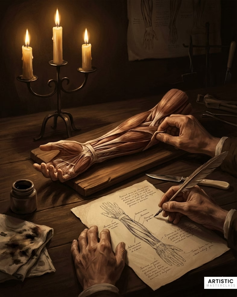
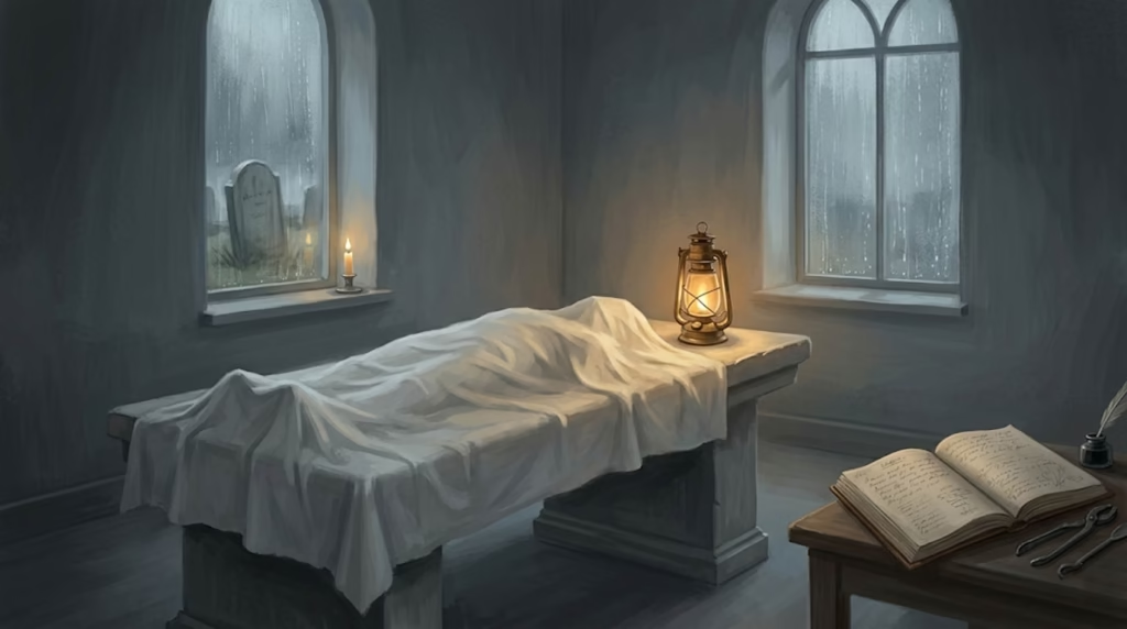
But serious artists went further. They arranged private dissections, sometimes conducting them personally. This meant confronting aspects of human mortality that most people never witnessed.
The process was difficult. Bodies decayed rapidly without refrigeration. The smell was overwhelming. The work had to be done quickly, often by candlelight, before putrefaction made observation impossible. Leonardo da Vinci himself wrote about “the fear of living through the night hours in the company of quartered and flayed corpses fearful to behold.”
The most dedicated artists created écorchés—carefully dissected bodies where successive layers of skin, fat, and muscle were peeled back to reveal the structures beneath. These weren’t just teaching tools; they were studies in form and structure that informed everything these artists created.
What Artists Brought to Anatomy
Anatomists working alone had made observations for centuries. But they struggled to communicate what they saw. Medical illustrations before the Renaissance were schematic at best, often misleading.
Artists brought something anatomists lacked: the ability to translate three-dimensional forms into clear two-dimensional representations. They understood perspective—how to show depth and spatial relationships on a flat surface. They knew how to use shading to suggest volume, line weight to indicate importance, and composition to guide the viewer’s eye.
More fundamentally, artists were trained observers. They noticed subtle curves, slight asymmetries, the way light revealed structure. Where a physician might focus on pathology or function, an artist saw form, proportion, and relationship.
When these skills combined with anatomical knowledge, the results were revolutionary.
Leonardo da Vinci: The Master of Both Worlds
No figure better exemplifies the Renaissance artist-anatomist than Leonardo da Vinci. His anatomical work represents not just artistic achievement but genuine scientific discovery.
Early Anatomical Studies (1480s-1490s)
Leonardo’s anatomical investigations began as many artists’ did—with the practical goal of improving his art. In the late 1480s, he started sketching from live models and cadavers, trying to understand the body’s proportions and mechanics.
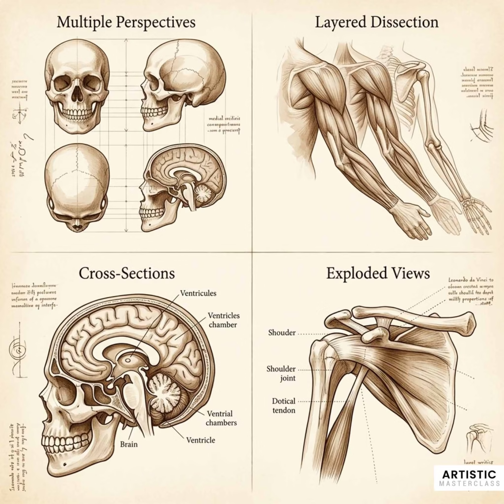
His 1489 skull studies show something new in anatomical illustration. Rather than a single view, Leonardo drew the skull from multiple angles—front, side, top, and in cross-section. He adopted techniques from architectural drawing, treating the skull as a complex structure to be mapped from every perspective.
These weren’t just artist’s sketches. Leonardo was beginning to ask scientific questions. How does the skull’s structure relate to its function? Where exactly do the nerves enter? What’s the relationship between outer form and inner cavity?
Collaboration with Marcantonio della Torre (1510-1511)
The most productive period of Leonardo’s anatomical work came around 1510-1511, when he collaborated with Marcantonio della Torre, a young professor of anatomy at the University of Pavia.
This partnership shows how artist-anatomist collaboration worked in practice. Della Torre provided access to cadavers and anatomical expertise, explaining what they were seeing and placing observations in the context of medical knowledge. Leonardo provided illustration skills and a way of seeing that led to genuinely new discoveries.
By his own count, Leonardo dissected approximately 30 human bodies during his lifetime. During this intensive period with della Torre, he created his most precise and scientifically valuable drawings—detailed studies of the heart, brain, respiratory system, and musculoskeletal structure.
Tragically, della Torre died of plague in 1511, ending the collaboration. Leonardo never found another partner who combined scientific knowledge with willingness to work with an artist.
Leonardo’s Innovations in Anatomical Illustration
Leonardo didn’t just draw what he saw—he invented new ways of representing anatomical information.
He pioneered the use of multiple perspectives, showing the same structure from front, side, and back views on the same page. This technique, borrowed from architectural drawing, allowed viewers to build a three-dimensional understanding from two-dimensional images.
He developed layered illustrations that showed progressive dissection. The first drawing might show skin and surface muscles, the second deeper muscles, the third bones and organs. This sequential approach taught viewers how to understand the body’s layered construction.
He created cross-sectional views, slicing through structures to reveal their internal organization. His cross-sections of the skull showing brain ventricles were unprecedented in their clarity and accuracy.
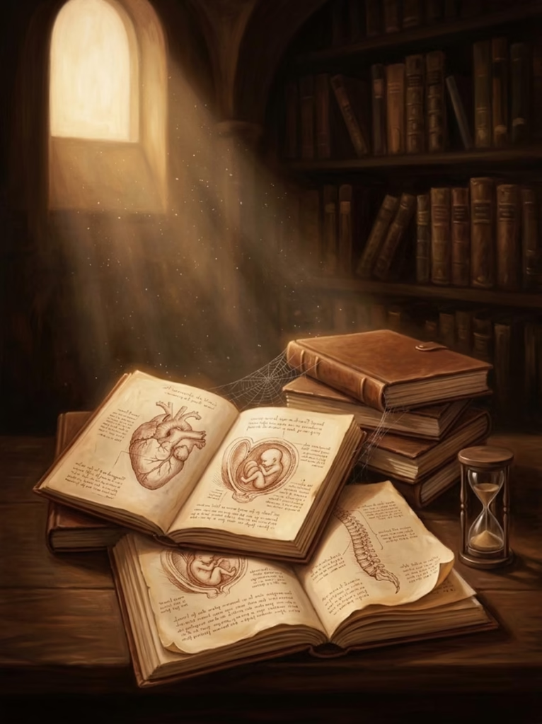
He made genuine discoveries. Leonardo produced the first accurate depiction of the human spine, correctly showing its curves. He documented the first known description of cirrhosis of the liver and atherosclerosis in the arteries. His drawings of a fetus in the womb, based on dissection of a pregnant woman who died in childbirth, were anatomically accurate and wouldn’t be equaled for centuries.
Most remarkably, he used what he called “exploded views”—drawings that separated structures slightly to show their relationships while maintaining proportional accuracy. This technique is still used in technical illustration today.
Why Leonardo’s Work Remained Hidden
Here’s the tragedy: almost none of this reached the people who needed it most.
Leonardo intended to publish a comprehensive treatise on human anatomy. His notebooks contain plans for the work, organized by body systems. But he never completed it. After his death in 1519, his notebooks were scattered among collectors and heirs. Many disappeared. Others remained in private collections, unseen by scientists or artists.
Some of Leonardo’s anatomical drawings weren’t rediscovered and published until the late 19th or early 20th century—400 years after they were created. By then, medicine had long since made the same discoveries independently.
This raises a fascinating question: how might medical history have been different if Leonardo’s work had been published in his lifetime? We’ll never know. But his innovations in illustration technique did gradually spread through artists’ networks and influenced the next generation of anatomical atlases, even if indirectly.
Artist-Anatomist Collaborations: A New Model of Knowledge Creation
Leonardo represents the ideal of a single individual mastering both art and science. But the more common pattern was collaboration between specialists—artists and anatomists working together, each contributing what the other lacked.
Michelangelo: Art Informed by Anatomy
Michelangelo Buonarroti approached anatomy from the opposite direction of Leonardo. Where Leonardo was motivated by scientific curiosity, Michelangelo sought anatomical knowledge purely to perfect his art.
He attended dissections and conducted his own anatomical studies, but his goal was always to capture the human form at its most powerful and expressive. The muscular dynamism of his figures in the Sistine Chapel ceiling shows deep understanding of how muscles layer over bone, how tendons connect to joints, how the body’s weight shifts in motion.
His David demonstrates anatomical accuracy in every detail—the tension in the neck muscles, the engagement of core muscles in the contrapposto stance, the veins visible in the hands. Yet it’s not a medical illustration. It’s anatomy in service of ideal beauty and expressive power.
Michelangelo even embedded anatomical references in his paintings. In the Last Judgment on the Sistine Chapel’s altar wall, Saint Bartholomew holds his own flayed skin—but the face on that skin appears to be Michelangelo’s self-portrait. It’s both a meditation on martyrdom and a display of anatomical knowledge, showing the artist’s familiarity with the écorché tradition.
Raphael and Berengario da Carpi
A recent scholarly discovery illuminates how artist drawings became scientific illustrations. Raphael, one of the Renaissance’s great masters, created anatomical sketches that found their way into Giacomo Berengario da Carpi’s anatomical treatise Commentaria (1521).
Berengario was both a physician-anatomist and an art collector with a particular passion for Raphael’s drawings. When he needed illustrations for his anatomical treatise, he turned to drawings in his collection. At least one of Raphael’s anatomical studies became the direct source for a printed plate in Berengario’s work.
Through that printed version, Raphael’s drawing influenced Andreas Vesalius’s later masterpiece De humani corporis fabrica. A sketch created for artistic purposes became part of the scientific canon, teaching medical students for generations.
This reveals how porous the boundaries were. Art collections, scientific treatises, and educational materials all drew from the same pool of anatomical knowledge. The distinctions we make today between “art” and “science” simply didn’t exist in the same way.
The Practical Reality of Partnership
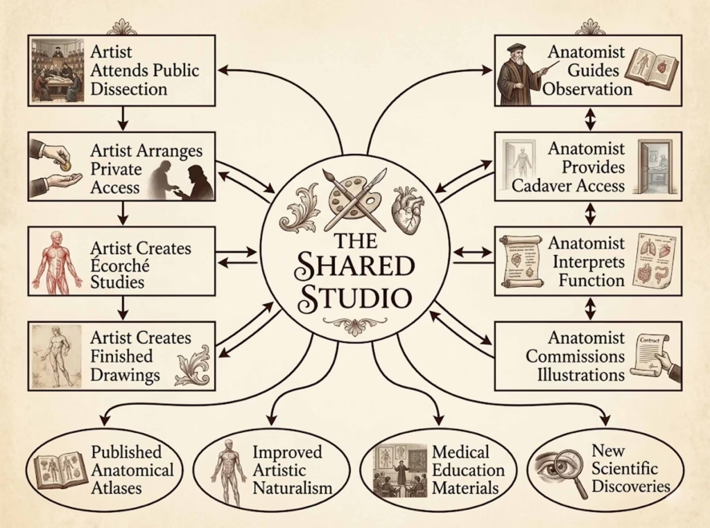
What did these collaborations actually look like day-to-day?
Artists were sometimes hired by anatomists specifically to create illustrations for medical texts. The contract would specify the number of plates, the payment, and sometimes even the artistic style expected. The anatomist would guide what to show, which structures to emphasize, what labels to include. The artist would determine how to compose the image, what perspective to use, how to render texture and depth.
In other cases, anatomists provided access to dissections in exchange for illustrations. An artist might attend multiple dissections of the same body part, making sketches and notes, then create finished drawings based on composite observations.
Some artists, like Leonardo, worked more independently but consulted with physicians to understand what they were seeing. The physician’s role was to interpret—explaining function, correcting misidentifications, placing structures in the broader context of bodily systems.
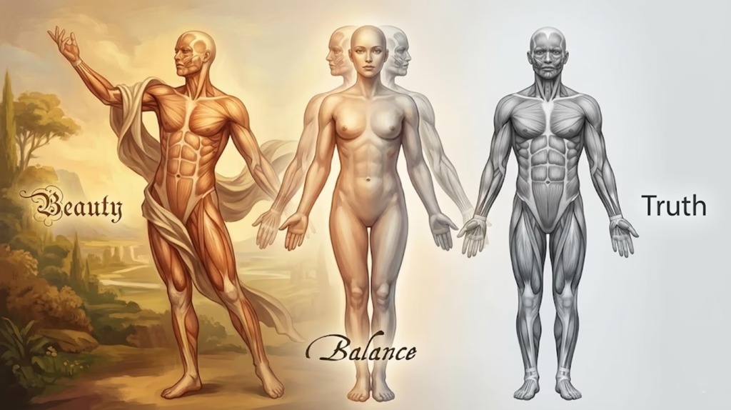
These weren’t always harmonious partnerships. Artists sometimes felt constrained by anatomists’ demands for accuracy over aesthetic appeal. Anatomists sometimes felt artists took too many liberties in the service of composition. But the tension was productive, forcing both parties to balance competing demands of truth and beauty.
Andreas Vesalius: When Science Met Sophisticated Art
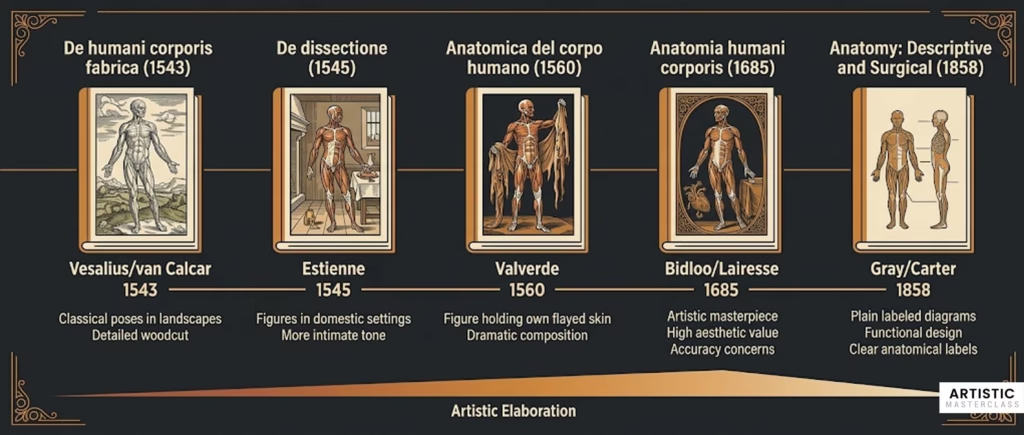
In 1543, a book was published that would change both medicine and anatomical illustration forever. Andreas Vesalius’s De humani corporis fabrica (On the Fabric of the Human Body) represented the pinnacle of Renaissance artist-anatomist collaboration.
Challenging Galen Through Direct Observation
Vesalius was a young professor of surgery and anatomy at the University of Padua. He had a radical conviction: Galen was wrong about human anatomy because Galen had never dissected a human body.
This was an extraordinarily bold claim. Galen’s authority had been unquestioned for 1,400 years. Challenging him was almost like challenging scripture.
But Vesalius had evidence. Through systematic dissection—he performed all his own dissections, unusual for a professor—he discovered error after error in Galenic anatomy. The human liver had only two lobes, not five. The human sternum had three segments, not seven. The interventricular septum of the heart was solid, not porous.
To prove these claims, Vesalius needed more than words. He needed illustrations so clear, so detailed, so obviously based on direct observation that they would compel belief. He needed an artist of exceptional skill.
The Artist Behind the Masterpiece
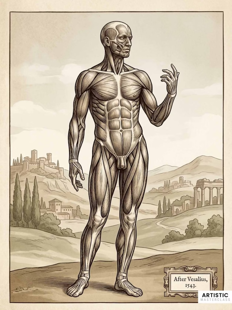
The illustrator of the Fabrica was likely Jan Stephen van Calcar, a student of the great Venetian painter Titian. The attribution isn’t certain—Vesalius never named his artist—but stylistic analysis and contemporary references point to van Calcar.
Whoever the artist was, they made choices that transformed anatomical illustration. Rather than placing dissected bodies on plain backgrounds or dissection tables, the figures stand in elaborately detailed Italian landscapes. Classical architecture frames the scenes. The dissected bodies are posed not as corpses but as living figures in dynamic stances.
A skeleton leans contemplatively against a tomb, its hand supporting its skull as if pondering mortality. A figure with its muscles exposed gestures dramatically against a backdrop of rolling hills. These are simultaneously scientific demonstrations and philosophical meditations on human existence.
The artistic decisions weren’t mere decoration—they served pedagogical purposes. The classical poses helped students understand how muscles functioned in life, not just how they appeared in death. The landscape backgrounds provided scale references. The dramatic compositions made the images memorable, aiding retention.
Production and Impact
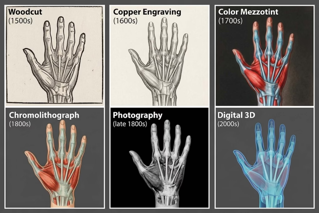
The Fabrica was printed in Basel in 1543 as a large folio volume with woodcut illustrations. The wood blocks were carved with extraordinary precision, capturing every detail of van Calcar’s drawings.
The publication was immediately recognized as revolutionary. Vesalius wasn’t just correcting Galen—he was establishing a new standard for anatomical study based on direct observation rather than ancient authority.
His book became the foundation for modern anatomy. Medical students for the next 200 years learned anatomy from Vesalius’s illustrations or from works directly based on them. The visual vocabulary he and his artist established—the poses, the perspectives, the way of rendering muscle and bone—became the standard approach.
But perhaps most importantly, the Fabrica demonstrated that anatomical illustration was essential to anatomical knowledge, not supplementary to it. You couldn’t fully understand anatomy from text alone. The image was the argument.
The Golden Age: 16th-17th Century Anatomical Publishing
Following Vesalius’s breakthrough, anatomical publishing exploded across Europe. Artists and anatomists experimented with different approaches, creating a rich diversity of illustrated anatomical works.
Variations on the Vesalian Theme
Charles Estienne’s De dissectione partium corporis humani (1545) took a different approach. His figures appeared in domestic settings—near fireplaces, leaning against doorframes—making anatomy feel less clinical and more integrated with everyday life.
Juan Valverde de Amusco’s Anatomica del corpo humano (1560) featured one of the most striking images in anatomical illustration history: a figure holding up its own flayed skin while standing in a classical pose. It was simultaneously horrifying and beautiful, a meditation on what lies beneath the surface of human identity.
Innovative formats emerged. “Flap anatomies” used layered paper cutouts that lifted to reveal progressively deeper structures. Readers could literally peel back the layers of the body, moving from skin to muscle to organs to skeleton. These interactive books made anatomy tangible in a new way.
Different national styles developed. Italian anatomists favored dramatic poses and classical references. Dutch anatomies emphasized precision and detail. French works often incorporated color to distinguish different structures.
When Art Overshadowed Accuracy
Not every collaboration succeeded in balancing artistic beauty with scientific accuracy. Govard Bidloo’s Anatomia humani corporis (1685), illustrated by the accomplished artist Gérard de Lairesse, shows the tensions that could arise.
Lairesse’s plates are magnificent works of art. The compositions are sophisticated, the rendering masterful, the overall effect visually stunning. But they contain numerous anatomical inaccuracies. Form triumphed over function, beauty over precision.
Anatomists criticized the work. Some, like William Cowper, literally stole the illustrations and republished them with corrected text. The controversy revealed growing anxiety about the proper relationship between art and science in anatomical illustration.
Should anatomical images be beautiful or merely accurate? Was artistic elaboration helpful for engaging students, or did it distract from the scientific content? These debates would intensify over the following centuries.
Innovation in Techniques
The 17th century saw constant technical innovation in illustration and printing methods.
Woodcuts gave way to copper plate engravings, which allowed finer detail and more subtle gradations of tone. Artists like Bidloo’s illustrator Lairesse exploited these capabilities to create images of unprecedented refinement.
Color printing began to emerge. Jacques Fabien Gautier d’Agoty pioneered color mezzotint techniques in the mid-18th century, creating anatomical plates in full color that revealed the body’s “horrible precision” in vivid reds, blues, and yellows. His works were as much art objects as medical references.
Publishers created different editions for different markets. Wealthy patrons bought large folio volumes with full-page illustrations on heavy paper. Medical students used smaller, cheaper editions with simpler illustrations. The same anatomical knowledge was packaged differently depending on the audience and purpose.
The 18th Century: Science Reasserts Control
By the 18th century, the relationship between art and science in anatomical illustration was shifting. Science was beginning to assert its independence from aesthetic concerns.
The Measured Precision of Albinus
Bernhard Siegfried Albinus, working at the University of Leiden in the 1730s-1770s, represented a new approach. He insisted on absolute accuracy, achieved through systematic measurement rather than artistic interpretation.
Albinus employed the artist Jan Wandelaar, but under strict supervision. Using grids, measuring instruments, and careful notation, they created composite figures based on observations of multiple cadavers. Rather than drawing what any single body looked like, they aimed to capture the ideal form, distilled from many examples.
The resulting plates combined scientific precision with undeniable artistic accomplishment. Skeletons posed beside rhinoceroses (after the famous Clara, who toured Europe as a curiosity). Muscle men stood in carefully rendered landscapes. These images were both more accurate than Vesalius and equally beautiful.
Albinus’s methods spread rapidly, particularly to Britain. His plates became the new standard, replacing Vesalius’s 200-year-old images as the definitive anatomical reference.
But note the shift: the artist was now clearly subordinate to the anatomist. Wandelaar was executing Albinus’s vision, not collaborating as an equal partner. The “shared studio” was becoming a hierarchical relationship.
William Hunter and Specialized Anatomy
William Hunter’s Anatomia uteri humani gravidi (1774), focused specifically on the pregnant uterus, showed anatomy becoming more specialized. Rather than comprehensive anatomies covering the whole body, increasingly detailed works examined specific organs or systems.
Hunter hired Jan van Rymsdyk to create life-size illustrations showing the uterus at different stages of pregnancy. These plates were startlingly realistic, even uncomfortable in their unflinching depiction of maternal and fetal structures.
The work represented a maturation of anatomical illustration. It was no longer enough to show basic structure. Advanced medical practice required detailed understanding of variations, developmental stages, and pathological conditions.
The Push for Realism Over Rhetoric
Between 1680 and 1800, a movement emerged among anatomists to strip away what they saw as unnecessary artistic rhetoric. They argued that metaphor, death imagery, and theatrical gestures didn’t belong in scientific illustration.
This “getting real” movement wanted objective representation—anatomy as it actually appeared on the dissection table, without artistic enhancement or philosophical commentary.
But even as they rejected overt artistry, these “realistic” illustrations developed their own aesthetic. They used intense color, sumptuous textures, and radical partitioning of the body. The “style-less” style was still a style—it just pretended not to be.
The 19th Century: The Parting of Ways
The final separation of art and science in anatomical illustration came in the 19th century. This shift fundamentally changed how anatomy was taught and visualized.
Gray’s Anatomy: Deliberately Style-Less
When Henry Gray’s Anatomy: Descriptive and Surgical was published in 1858, it represented a conscious rejection of artistic elaboration in favor of pure functionality.
Gray and his illustrator, Henry Vandyke Carter, had a specific goal: create a practical, affordable anatomy textbook for medical students. They wanted students to feel “as close to the dissecting table as possible without physically witnessing” dissection.
Carter’s woodcut illustrations were deliberately plain. No landscapes. No classical poses. No artistic flourishes. Just labeled anatomical structures rendered with sufficient detail to support surgical training.
The labels themselves became integral to the illustrations—not afterthoughts but essential elements of the image. This approach, which seems obvious to us now, was innovative in 1858.
Gray’s Anatomy was compact (compared to earlier folio volumes), relatively inexpensive, and eminently practical. It became the standard anatomical text for English-speaking medical students and has remained in print, continuously updated, for over 165 years.
But something was lost. Carter’s “style-less” illustrations lacked the visual drama, the philosophical depth, and the sheer beauty of Renaissance and Baroque anatomies. They were tools, not art objects.
Why the Change Happened
Multiple factors drove this transformation:
Medical schools were proliferating, creating demand for practical, affordable texts. The grand folio volumes of earlier centuries couldn’t meet this need.
Medicine was professionalizing, separating itself from the humanities and asserting its identity as a rigorous science. Scientific objectivity seemed to require rejecting artistic embellishment.
New printing technologies made mass production economically viable. Chromolithography could reproduce color illustrations cheaply. Smaller formats became standard.
The audience changed. Renaissance anatomies addressed wealthy patrons and educated gentlemen. Nineteenth-century texts targeted working medical students who needed practical knowledge, not philosophical meditation.
Perhaps most fundamentally, the unified worldview that had sustained artist-anatomist collaboration was fragmenting. Art and science were becoming separate domains with different values, methods, and goals.
New Technologies
The 19th century brought revolutionary technologies that began to supplement hand-drawn illustration:
Photography, invented in the 1830s-40s, initially couldn’t capture the detail needed for anatomical instruction. But it improved rapidly and by century’s end was being used to photograph dissections and anatomical preparations.
Frozen cross-sections, pioneered by de Riemer, allowed anatomists to see the body’s internal organization in situ. Corpses were frozen solid, then sawed into slices. Photographing these sections created anatomical references that drawing couldn’t match.
Wax modeling created three-dimensional teaching tools. Artists sculptured anatomically accurate models that could be handled, rotated, and studied from all angles—something impossible with two-dimensional illustrations.
These technologies didn’t replace illustration immediately. But they pointed toward a future where anatomical knowledge would be captured through mechanical and photographic means rather than artist’s interpretation.
The Dark Side: Ethics and Body Procurement
The history of anatomical illustration has a shadow side that’s essential to understanding the full story. The beautiful drawings we admire today were made possible by a morally troubling system of body procurement.
The Shortage of Legal Cadavers
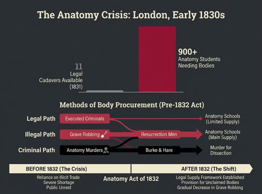
Throughout most of this history, human dissection was restricted to executed criminals. The logic was that those who had broken society’s laws had forfeited their right to bodily integrity after death.
But the supply was grotesquely inadequate. In 1831 London, for example, only 11 bodies were legally available for dissection—to serve over 900 anatomy students. The mathematics simply didn’t work.
Medical schools and anatomists faced a choice: limit anatomical education severely, or find bodies through other means. Most chose the latter.
Grave Robbing and Body Snatching
“Resurrection men” became a recognized profession. These body snatchers would rob fresh graves, stealing corpses to sell to anatomists and teaching hospitals.
The trade was lucrative. A fresh body could fetch several pounds—a substantial sum for working-class resurrection men. Some anatomists maintained regular business relationships with body snatchers, asking no questions about how cadavers were obtained.
There was a clear class dimension to this practice. Wealthy people could afford secure graves—brick-lined vaults, locked crypts, employed grave watchers. The poor, buried in paupers’ graves or common plots, had no such protection. Their bodies were vulnerable.
Artists and anatomists who benefited from this system were morally complicit in it, even if they didn’t personally rob graves. The beautiful anatomical drawings we admire were often based on bodies stolen from grieving families.
Anatomy Murders
The most horrifying development was murder-to-order. The notorious case of William Burke and William Hare in Edinburgh (1828) revealed that some people would kill to supply the anatomy trade.
Burke and Hare murdered 16 people, selling their bodies to anatomist Robert Knox. They targeted vulnerable individuals—the poor, the friendless, those who wouldn’t be missed. When the crimes were discovered, public outrage forced reform.
Anatomy Acts and Reform
England’s Anatomy Act of 1832 attempted to address the crisis by making unclaimed bodies—those of people who died in workhouses, prisons, or asylums—available for dissection.
This solved the supply problem but institutionalized class bias. Wealthy people’s bodies remained protected. The poor, the institutionalized, and the marginalized became the raw material for anatomical education.
In America, the bodies of enslaved people were used for dissection without consent and often without knowledge of their families. This exploitation continued after emancipation, with Black bodies disproportionately targeted for anatomical study well into the 20th century.
The ethical issues raised by this history remain relevant today. Modern anatomical study relies on body donation programs and explicit consent. But we should acknowledge that the foundations of anatomical knowledge were built, in part, on bodies taken without consent from the vulnerable.
Legacy: How the Shared Studio Shaped Modern Medicine and Art
The Renaissance collaboration between artists and scientists left lasting marks on both fields. Understanding this legacy helps us see how that brief “shared studio” moment continues to influence how we visualize and understand the human body.
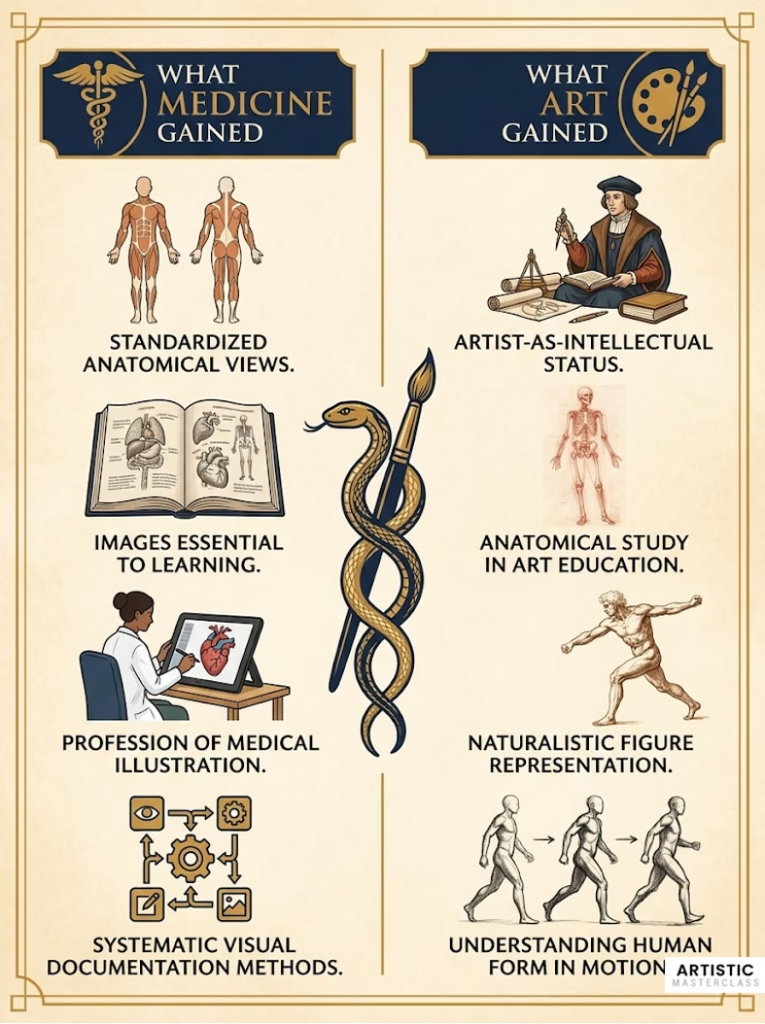
What Medicine Gained
The most obvious legacy is a visual vocabulary. The conventions established by Renaissance artist-anatomists—the standard views, the use of labels, the ways of rendering different tissues—still structure medical illustration today.
Medical education internalized the understanding that accurate representation is essential for learning anatomy. You can’t train surgeons with text alone. The image isn’t supplementary to knowledge—it is knowledge in a form that can’t be reduced to words.
Modern medical illustration remains a specialized field requiring both artistic skill and anatomical knowledge. Medical illustrators study anatomy as intensively as medical students while mastering drawing, painting, digital illustration, and 3D modeling. The Renaissance ideal of the artist-anatomist persists in this profession.
What Art Gained
Artists’ status as intellectuals, not mere craftsmen, was cemented partly through their engagement with anatomy. The Renaissance establishment of drawing from life and from dissection became fundamental to art education.
Even today, serious art training includes anatomical study. Students at major art schools sketch skeletons, learn muscle groups, and study the body’s structure and mechanics. The knowledge Leonardo and Michelangelo sought is still considered essential for figure drawing and sculpture.
Renaissance anatomical knowledge enabled a revolution in naturalistic representation. The ability to depict human figures in complex poses, in motion, in foreshortened perspectives, with accurate musculature—all of this flows from the anatomical study pioneered during this period.
The Second Renaissance: Contemporary Convergence
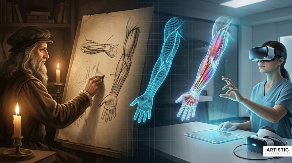
Interestingly, we’re seeing a new convergence of art and science in anatomical visualization. Digital technology is creating a “second Renaissance” where creative and technical skills again combine.
Medical illustrators use 3D modeling software to create virtual anatomies that can be rotated, sectioned, and explored interactively. Virtual reality allows medical students to “dissect” digital cadavers, combining the visual richness of traditional anatomical art with interactive capabilities impossible in static images.
Artists continue to engage with anatomy in contemporary work. Body painters create elaborate anatomical designs on living skin, teaching surface anatomy in visually striking ways. Artists like Damien Hirst incorporate actual anatomical specimens in works that blur boundaries between art, science, and spectacle.
Some medical schools are even returning to Renaissance art to teach anatomy. Students analyze Michelangelo’s paintings and Leonardo’s drawings to understand musculoskeletal anatomy and surface landmarks. The 500-year-old collaboration still has pedagogical value.
The tools have changed—from charcoal and dissection tables to 3D scanners and VR headsets—but the fundamental insight remains: understanding the human body requires both scientific precision and visual intelligence. The studio that artists and scientists shared during the Renaissance continues to exist, now in digital space.
Frequently Asked Questions About Anatomical Drawing History
Why did Renaissance artists study anatomy?
Renaissance artists studied anatomy to create more naturalistic, lifelike artwork depicting human figures in accurate proportions and dynamic poses. Under the influence of humanism, which emphasized the beauty and importance of the human form, artists needed to understand not just surface appearance but underlying musculoskeletal structure. This knowledge allowed them to paint and sculpt figures in complex positions, in motion, and with anatomical accuracy that distinguished Renaissance art from earlier medieval work.
How did Leonardo da Vinci study anatomy?
Leonardo studied anatomy through direct dissection of approximately 30 human cadavers over two decades, often working at night by candlelight in hospitals and morgues. He collaborated with physician-anatomist Marcantonio della Torre at the University of Pavia around 1510-1511, combining his artistic observation skills with medical expertise. Leonardo pioneered innovative illustration techniques including multiple perspective views, cross-sections, and layered drawings showing progressive dissection. His anatomical studies led to genuine scientific discoveries, including the first accurate depiction of the human spine and the earliest known description of atherosclerosis.
What was the first illustrated anatomical book?
The Fasciculus Medicinae (1491) was the first printed medical text with significant anatomical illustrations, though these were still medieval in character. However, Andreas Vesalius’s De humani corporis fabrica (1543) was the first comprehensive anatomical atlas with scientifically accurate illustrations based on systematic human dissection. Vesalius’s work revolutionized anatomical illustration by combining scientific precision with sophisticated artistic rendering, establishing the foundation for modern anatomical publishing.
Who illustrated Andreas Vesalius’s anatomy book?
The illustrator was likely Jan Stephen van Calcar, a student of the great Venetian painter Titian, though Vesalius never definitively named his artist. Stylistic analysis and contemporary references point to van Calcar as the most probable creator of the dramatic woodcut illustrations. These images showed dissected bodies posed in classical stances within elaborate Italian landscapes, combining scientific demonstration with philosophical meditation on human nature.
Why did anatomical illustration become less artistic over time?
Several factors drove this shift in the 18th and 19th centuries. Medical professionalization led physicians to distinguish their scientific discipline from the humanities, making artistic embellishment seem inappropriate. The growth of medical schools created demand for practical, affordable textbooks rather than expensive art objects. New printing technologies enabled mass production at lower cost. Additionally, changing educational philosophy emphasized functional clarity over aesthetic beauty—medical students needed practical guides to dissection, not philosophical meditations on mortality. This culminated in works like Gray’s Anatomy (1858), which deliberately adopted a “style-less” approach focused purely on anatomical instruction.
What are écorchés?
Écorchés (from French “flayed”) are anatomical studies of dissected bodies with skin and successive layers removed to reveal underlying muscles, tendons, and bones. Renaissance artists and anatomists created écorchés to understand the body’s layered structure and how muscles functioned beneath the skin. These ranged from drawings showing progressive dissection to actual cadavers carefully preserved with different layers exposed. The écorché tradition influenced both artistic training (teaching how muscles create surface form) and anatomical education (demonstrating muscle origins, insertions, and relationships).
How did artists get access to cadavers for study?
Artists obtained cadavers through several methods, though with varying legality. Some attended public dissections held at university anatomical theaters in cities like Padua and Bologna. More serious students arranged private access through relationships with physician-anatomists, sometimes assisting with dissections in exchange for learning opportunities. A few artists conducted their own dissections, though this required either official permission or willingness to work secretly. The scarcity of legal cadavers (restricted to executed criminals) meant that both artists and anatomists sometimes relied on bodies obtained through grave robbing or other questionable means.
What role did the printing press play in anatomical illustration?
The printing press, perfected around 1450, revolutionized anatomical knowledge dissemination. For the first time, identical anatomical illustrations could be reproduced across hundreds or thousands of copies, standardizing visual references across Europe. This enabled Vesalius’s De humani corporis fabrica to establish new anatomical knowledge broadly and rapidly. The printing press also made illustrated anatomical texts economically viable, creating a market for anatomical publishing that drove innovation in both scientific content and illustration techniques. Without printing, anatomical knowledge would have remained limited to hand-copied manuscripts with inevitable copying errors and variations.
What was Gray’s Anatomy?
Gray’s Anatomy (formally Anatomy: Descriptive and Surgical) was an anatomical textbook first published in 1858 by English anatomist Henry Gray with illustrations by Henry Vandyke Carter. It revolutionized medical education by providing a compact, affordable, deliberately functional anatomy text for medical students. Carter’s illustrations rejected artistic embellishment in favor of clear, labeled diagrams designed to support surgical training. The book’s practical approach made it enormously successful; it remains in print today after 42 editions spanning 167 years, continually updated to reflect current anatomical knowledge.
How did religious beliefs affect anatomical study during the Renaissance?
Both Christian and Islamic traditions initially viewed dissection as desecration of the sacred human body. In medieval Europe, the Church’s theological position that bodies would be resurrected made cutting them problematic. However, restrictions gradually relaxed during the Renaissance. Papal decrees allowed limited dissection of executed criminals for medical education at universities. Islamic scholars faced similar restrictions under Quranic law but nonetheless preserved classical anatomical texts and made contributions through careful observation and animal dissection. The Renaissance humanist movement, while still Christian, emphasized understanding God’s creation through direct study of nature, including the human body, creating philosophical space for anatomical investigation.
Key Takeaways
The history of anatomical drawing reveals that great scientific and artistic achievements often emerge from collaboration, not isolation. When Renaissance artists and anatomists shared studios and dissection rooms, they created something neither could have achieved alone—a visual language for understanding the human body that remains foundational to both medicine and art.
The cultural conditions that enabled this collaboration—humanism’s emphasis on the human form, wealthy patronage supporting both disciplines, the printing press enabling knowledge dissemination, and the gradual relaxation of religious restrictions—remind us that scientific and artistic progress depend on broader social and philosophical contexts.
Leonardo da Vinci’s anatomical work exemplifies the power of combining artistic observation with scientific inquiry, making discoveries through visual thinking that pure anatomy might have missed. Yet the tragedy of his unpublished notebooks shows that even brilliant work has little impact if it can’t be shared.
The gradual separation of art and science in the 18th and 19th centuries, culminating in deliberately “style-less” medical illustration, raises questions about what was gained and lost. We achieved greater standardization and accessibility, but perhaps sacrificed the philosophical depth and visual engagement that made Renaissance anatomies transformative.
Most importantly, this history teaches us that our current distinctions between artistic and scientific ways of knowing aren’t inevitable. They’re historically contingent. As digital technology creates new possibilities for anatomical visualization, we’re seeing a “second Renaissance” where creative and scientific skills again converge—suggesting that the shared studio Leonardo and Vesalius knew might have a future as well as a past.
For an even fuller account, see our complete history of anatomical drawing from ancient medicine to digital art, plus the related history of the self-portrait — both chapters in the evolution of art.